Meatus acusticus internus WikiSkripta
Meatus acusticus internus 1/6. Synonyms: Internal auditory canal, Canalis auditorius internus Clinical note Jugular foramen syndrome. Jugular foramen syndrome (JFS), also known as Vernet's syndrome is a disorder involving the palsies of the glossopharyngeal, vagus and accessory nerves (cranial nerves IX-XI), as well as sometimes the hypoglossal.

Meatus acusticus internus Ars Neurochirurgica
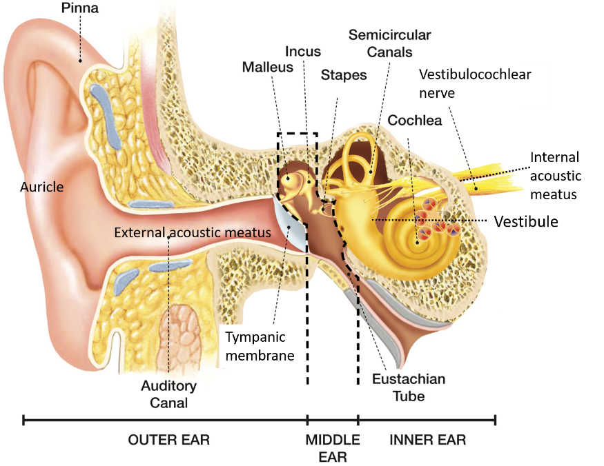
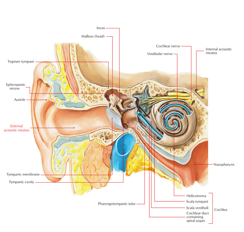
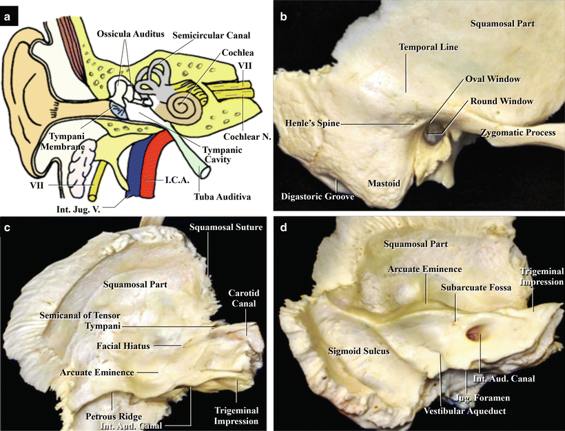
The internal auditory canal (IAC), also referred to as the internal acoustic meatus lies in the temporal bone and exists between the inner ear and posterior cranial fossa. It includes the vestibulocochlear nerve (CN VIII), facial nerve (CN VII), the labyrinthine artery, and the vestibular ganglion.
External Ear , Auricle and External acoustic meatus , Anatomy QA
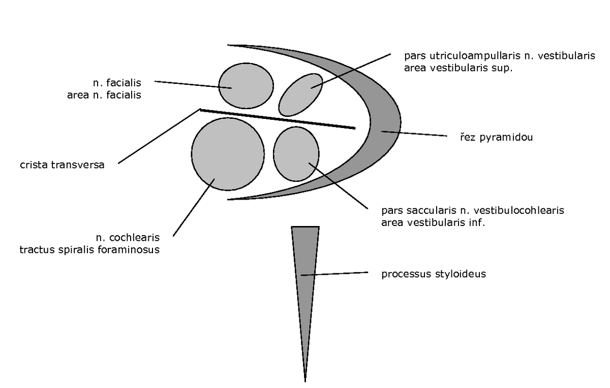
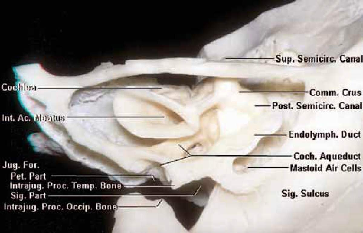
Definition The internal acoustic opening is a large orifice near the center of posterior surface of petrous part, its size varies considerably; its margins are smooth and rounded, and it leads into a short canal, the internal acoustic meatus, about 1 cm. in length, which runs lateralward.
Innerer Gehörgang Anatomie und Klinik Kenhub
Meatus acusticus can refer to: Ear canal (meatus acusticus externus) Internal auditory meatus (meatus acusticus internus) This disambiguation page lists articles associated with the title Meatus acusticus. If an internal link led you here, you may wish to change the link to point directly to the intended article.

10 hearing08
Internal ear This mixture of bones, nerves, vessels, membranes, and muscles that make up the ear will be described in this article. Contents External ear Auricle External acoustic meatus Tympanic membrane Muscles of the external ear Vasculature of the external ear Innervation of the external ear Middle ear Tympanic cavity Auditory ossicles
PPT Anatomy of the Ear PowerPoint Presentation ID6102402
The influences of porus acusticus internus on ethnicity and importance in preoperative and intraoperative approaches. R. Şekerci Eren Ogut N. Keles-Celik. Medicine.. Amac: Bu calisma 18-60 yas arasi saglikli Turk populasyonunda meatus acusticus internus morfometrisini belirlemek amaclandi, yurutulen bu calisma retrospektif bir calismadir.
Innerer Gehörgang Anatomie und Klinik Kenhub
The ear canal ( external acoustic meatus, external auditory meatus, EAM) is a pathway running from the outer ear to the middle ear. The adult human ear canal extends from the pinna to the eardrum and is about 2.5 centimetres (1 in) in length and 0.7 centimetres (0.3 in) in diameter. Structure The human ear canal is divided into two parts.
External Auditory Meatus/Acoustic Meatus Earth's Lab
The internal auditory meatus (IAM) is a small, bony canal located within the petrous portion of the temporal bone of the skull. It serves as an important pathway for various cranial nerves and blood vessels to pass through the inner ear.
external acoustic meatus bone location
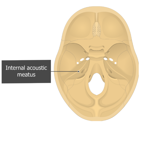
The internal auditory meatus (also meatus acusticus internus, internal acoustic meatus, internal auditory canal, or internal acoustic canal) is a canal within the petrous part of the temporal bone of the skull between the posterior cranial fossa and the inner ear . Structure
Internal Acoustic Meatus
The internal acoustic meatus was evaluated in 97 temporal bone specimens, half of which were radiographed in different projections.. Die Varianten des Meatus acusticus internus. Radiol. Diagn. 7 (1966), 141. PubMed. Google Scholar. 5. Camp J., Cilley.: The significance of asymmetry of the pori acustici as an aid in diagnosis of eighth nerve.
Oído medio Anatomía, partes, funciones Kenhub
Definition noun A short, narrow passageway through the temporal bone of the skull where the vestibular nerve and cochlear nerve pass through to reach the brainstem from the inner ear. Supplement In humans, the internal acoustic meatus is about 1-2 cm in length.
Temporal Bone Anatomy
Der Meatus acusticus internus ist ein kleiner, relativ kurzer Knochenkanal, der im Felsenbein (Pars petrosa ossis temporalis) verläuft. Er beginnt am Porus acusticus internus und endet an einer durchlöcherten Knochenplatte, die an das Innenohr grenzt. Anatomie.
external acoustic meatus bone location
Özocak O, Unur E, Ülger H, Ekinci N, Aycan K, Acer N. Meatus acusticus internus'un morfometrisi ve varyasyonları. Erciyes Üniversitesi Sağlık Bilimleri Dergisi. 2004;13(3) 1-7. 4. Kobayashi H, Zusho H. Measurements of internal auditory meatus by polytomography. The British Journal of Radiology 1987; 60 (711): 209-14.

External ear anatomy The pinna (A), the external acoustic meatus and... Download Scientific
Internal acoustic meatus refers to a small bony foramen situated on the posterior surface of the petrous part of temporal bone, inside the posterior cranial fossa. It allows for the passage of three important structures, namely the vestibulocochlear nerve, facial nerve and the labyrinthine artery.
PPT Anatomy of the Ear PowerPoint Presentation ID6102402
The porus acusticus internus (plural: pori acustici interni), often merely referred to as porus acusticus, is the medial opening of the internal acoustic canal through which the facial nerve , vestibulocochlear nerve and labyrinthine artery pass 1 .

Odyoloji Okuyoruz Biz
The internal auditory canal (IAC), also referred to as the internal acoustic meatus lies in the temporal bone and exists between the inner ear and posterior cranial fossa. It includes the vestibulocochlear nerve (CN VIII), facial nerve (CN VII), the labyrinthine artery, and the vestibular ganglion..
